Is Your Incision Infected or Just Healing? Redness vs. Cellulitis, Dermabond Glue Rash, and Why You Must Never Pull a Spitting Stitch
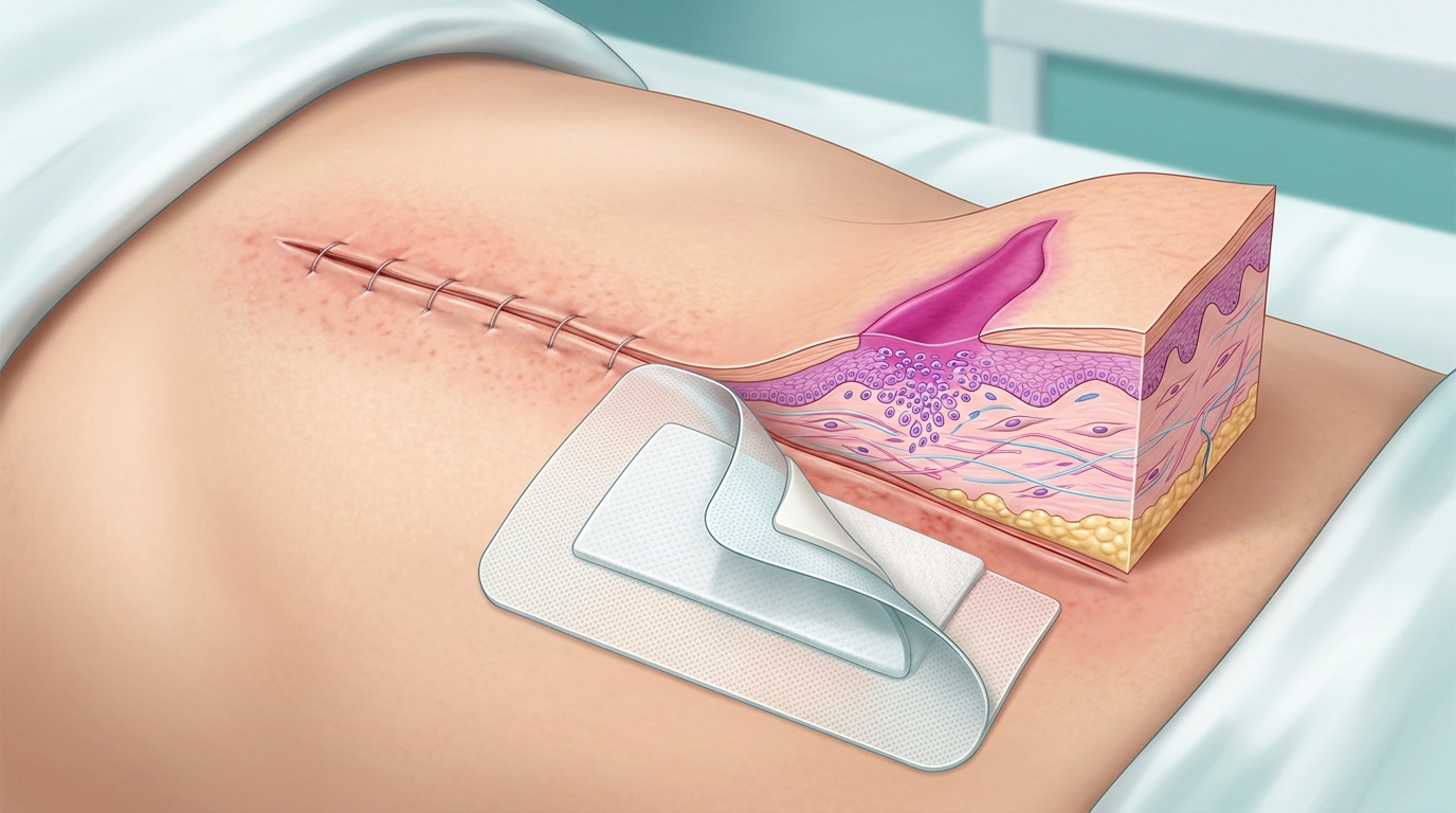
Normal healing redness is pale pink and tightly confined within 1 to 2 millimeters of your incision line. In contrast, a Surgical Site Infection (cellulitis) causes fiery redness spreading more than 1 to 2 centimeters away from the cut, accompanied by intense localized skin heat, wooden firmness (induration), throbbing pain, or cloudy foul-smelling pus. If an absorbable suture knot pokes out like a white pimple (a spitting stitch), never pull it with tweezers; wash gently with soap and water and let your surgeon snip it flush.
The Bathroom Mirror Inspection: Normal Hyperemia vs. True Infection
Every morning following outpatient surgery, patients stand in front of their bathroom mirror, gently lift their shirt, and scrutinize their surgical trocar incisions. Is that pink border normal? Why is it warm? Is that tiny drop of yellow fluid an infection, or is it normal healing?
According to Centers for Disease Control and Prevention (CDC) surveillance data, Surgical Site Infections (SSIs) occur in approximately 1% to 3% of clean laparoscopic operations [PMID: 28467526]. That means in 97% or more of cases, post-operative redness is benign physiological inflammation or a harmless allergic reaction to topical skin prep or surgical adhesives. Understanding how human skin regenerates—and how to perform the surgical pen marker test—will save you unnecessary panic and emergency room visits.
The Three Biological Phases of Incision Healing
When a surgeon closes a laparoscopic port or surgical incision using subcuticular sutures, surgical staples, or 2-octyl cyanoacrylate liquid glue (Dermabond), the anatomical repair progresses through three overlapping cellular phases:
- Phase 1: The Vascular & Inflammatory Phase (Hours 0 to 72): Injured platelets degranulate, releasing histamine, kinins, and prostaglandins. These chemical mediators cause localized arteriolar vasodilation. Capillaries become hyper-permeable, delivering neutrophils and monocytes to clear cellular debris and bacterial contaminants. This localized hypervascularity creates mild erythema (pinkness), faint warmth, and slight swelling tightly hugging the incision cut edge. This is NOT an infection; it is your immune system actively sealing the barrier.
- Phase 2: The Proliferative & Granulation Phase (Days 4 to 21): Vascular endothelial growth factor (VEGF) stimulates angiogenesis (new microscopic capillary loops). Fibroblasts synthesize an extracellular matrix of glycosaminoglycans and immature Type III collagen. In clean, approximated incisions, basal epidermal keratinocytes migrate across the wound margins, achieving complete surface re-epithelialization within 48 hours [PMID: 28368694]. This creates an impermeable biological seal against outside bathwater and environmental bacteria.
- Phase 3: The Maturation & Remodeling Phase (Weeks 3 to 1 Year): Disorganized collagen bundles are degraded by matrix metalloproteinases and replaced by dense, parallel Type I collagen fibers aligned along lines of biomechanical tension. The scar, which was initially raised, pink, and firm, slowly flattens, softens, and fades to a pale silver-white line over 6 to 12 months.
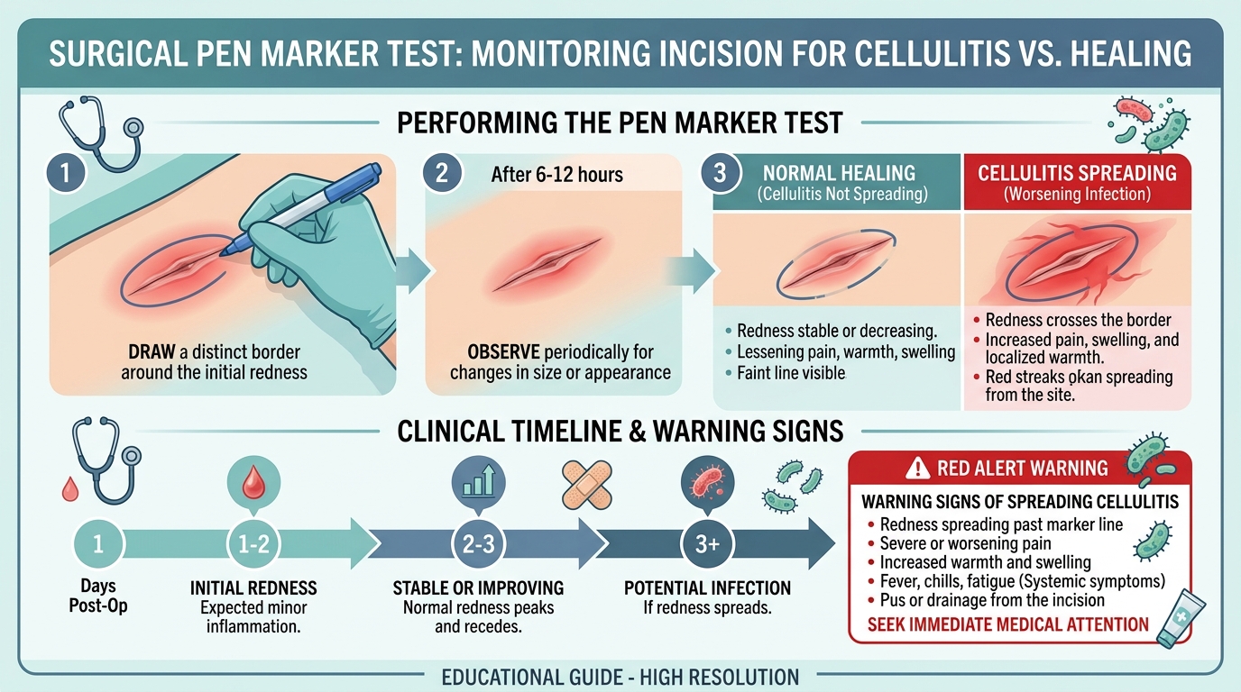
Figure 1: The Surgeon-Approved Pen Marker Test to definitively monitor spreading cellulitis versus benign physiological healing.
How to Perform the Surgical Pen Marker Test
If you notice redness around your laparoscopic port site or incision and cannot tell if it is expanding, perform the exact diagnostic test general surgeons use in hospital wards (Figure 1):
- Step 1 (Draw the Border): Take a clean, ballpoint pen or non-toxic fine-tip surgical marker. Trace a thin, distinct line directly along the outer perimeter of the redness.
- Step 2 (Periodic Observation): Do not touch, scrub, or apply unprescribed antibiotic ointments to the area. Go about your normal recovery day. Re-examine the incision in 6 to 12 hours under bright, direct light.
- Step 3 (Clinical Interpretation):
- NORMAL HEALING (Cellulitis Not Spreading): The redness remains strictly inside your pen line or begins to fade from bright red to pale pink. Local tenderness is decreasing. This confirms benign inflammatory hyperemia.
- CELLULITIS SPREADING (Worsening Infection): The fiery redness has crossed past your pen line, expanding outward into surrounding healthy skin. The skin feels hot to touch, firm like wood (induration), and throbbing. Call your surgeon immediately.
Clinical Differential: Normal Healing vs. Cellulitis vs. Dermabond Allergy
| Diagnostic Parameter | Normal Physiological Healing | Surgical Site Infection (Cellulitis/Abscess) | Allergic Contact Dermatitis (Dermabond/Tape) |
|---|---|---|---|
| Redness Margins & Shape | Light pink line confined tightly within 1–2 mm of the cut edge. | Fiery, angry red erythema spreading >1–2 cm away; irregular expanding borders. | Erythema matches the exact geometric shape of the adhesive tape or glue droplet. |
| Skin Temp & Palpation | Body temperature or faint warmth; soft, pliable surrounding tissue. | Intensely hot to touch; induration (tissue feels hard, wooden, and swollen). | Normal skin temperature; soft skin surface covered in tiny, itchy micro-blisters. |
| Dominant Sensation | Mild, decreasing tenderness that improves every 24 hours. | Deep, throbbing, escalating pain that worsens despite scheduled pain relievers. | Mild burning; primary symptom is intolerable, relentless cutaneous itching. |
| Wound Drainage (Exudate) | Completely dry, or minimal drops of clear amber (serous) or pink fluid in first 48 hrs. | Thick, opaque, creamy yellow or green pus; frequently possesses a foul, sour odor. | Clear, thin watery exudate only if superficial allergic blisters are scratched open. |
| Systemic Vital Signs | Completely normal; patient is afebrile with normal energy. | Oral temperature >100.4°F (38.0°C), shaking chills, malaise, tachycardia. | Completely normal; zero fever or systemic inflammatory symptoms. |
| Required Action | Keep clean and dry; gentle shower; no ointments needed. | Contact surgeon immediately; oral antibiotics (e.g., cephalexin) or suture removal to drain. | Gently remove loose glue with mineral oil/petroleum jelly; topical hydrocortisone if approved. |
What Is a "Spitting Suture"? (And Why You Must Never Pull It!)
Around Week 3, 4, or 6 post-surgery, patients frequently notice a tiny, raised, tender white pimple emerging directly along their surgical scar. A few days later, a clear, white, or dyed purple plastic thread pokes out through the center of the bump.
This is known clinically as a "spitting suture" (foreign body granulomatous extrusion) [PMID: 20706071].
Why Does It Happen?
Surgeons close deep dermal and subcuticular layers using synthetic absorbable sutures—such as polyglactin 910 (Vicryl), poliglecaprone 25 (Monocryl), or polydioxanone (PDS). These materials are engineered to dissolve inside living tissue via sterile chemical hydrolysis over 60 to 120 days.
However, if a suture knot was placed slightly too superficial in the dermis, or if your individual biological enzyme turnover is slow, your immune system recognizes the foreign synthetic polymer. White blood cells wall off the knot in a sterile granuloma and physically push it upward toward the skin surface to expel it.
The Golden Surgical Rule: NEVER Pull It With Tweezers!
It is immensely tempting to grab bathroom tweezers and pull on the exposed thread. Doing so is dangerous for three reasons:
- Unraveling Deep Structural Knots: The visible thread is tied to deep knots. Pulling on it can untie the knot before biological fascial integration is complete, triggering acute wound dehiscence.
- Tearing Deep Tissue: Forcing a knot through healing skin tears fragile collagen and multiplies your risk of an unsightly, widened scar.
- Seeding Skin Flora: As the thread pulls out, skin bacteria (like Staphylococcus aureus) are dragged into the sterile preperitoneal or deep subcutaneous pocket, transforming a harmless suture reaction into a deep surgical abscess!
What You Should Do Instead: Wash the area gently with antibacterial soap and warm water. Apply a clean adhesive bandage. Call your surgeon’s office. During a 30-second clinic visit, your surgeon will use sterile micro-scissors to snip the suture flush with the skin level, allowing the skin to close cleanly.
The Rules of Bathing: Showering vs. Submerging in Water
- Showering (Safe at 24–48 Hours): If your incisions are sealed with 2-octyl cyanoacrylate (Dermabond) or subcuticular stitches with waterproof Steri-Strips, you may take a warm, gentle shower after 24 to 48 hours. Let warm soapy water cascade passively over your incisions. Do NOT scrub, rub with washcloths, or direct high-pressure shower heads onto the wounds. Afterward, pat the incisions completely dry with a fresh, clean towel or let them air-dry.
- Submerging in Water (Strictly Prohibited for 3–4 Weeks): Never soak in bathtubs, hot tubs, swimming pools, lakes, or oceans until your surgeon formally clears you. Submerging wounds causes keratin maceration (softening and breakdown of the skin barrier) and introduces opportunistic environmental pathogens (such as Pseudomonas aeruginosa and aquatic atypical mycobacteria) [PMID: 28467526].
- Steri-Strips and Glue Care: Do not aggressively pick or peel surgical glue or paper tapes. Allow the edges to curl and lift naturally. By Day 10 to 14, any remaining loose edges can be gently loosened with warm water or a cotton swab dipped in mineral oil.
Scar Optimization: Silicone Gel Sheets vs. Vitamin E Myths
Once your incisions have fully epithelialized and any scabs have detached (typically between Weeks 3 and 4), patients want to know how to minimize their scars.
- The Proven Medical Standard: Medical-Grade Silicone Gel & Sheets. Decades of clinical trials in plastic and general surgery demonstrate that topical silicone creates an occlusive, semi-permeable hydration barrier that downregulates fibroblast collagen overproduction, preventing hypertrophic and keloid scars [PMID: 11594966]. Apply silicone sheets or gel twice daily for 8 to 12 weeks.
- UV Sun Protection (SPF 50+): Fresh scars contain hyperactive melanocytes. Direct sun exposure during the first 12 months causes permanent, dark hyperpigmentation (tanning of the scar). Keep scars covered or apply broad-spectrum mineral zinc oxide sunscreen.
- The Vitamin E Myth: Clinical trials show that applying topical Vitamin E capsules does NOT improve surgical scar cosmetic appearance and causes contact dermatitis in up to 30% of patients [PMID: 10417589]. Avoid it.
Peer-Reviewed Scientific References & Clinical Guidelines
- Berríos-Torres, S. I., et al. (CDC HICPAC). (2017). Centers for Disease Control and Prevention Guideline for the Prevention of Surgical Site Infection. JAMA Surgery, 152(8), 784–791. [PubMed PMID: 28467526].
- World Health Organization (WHO). (2016). Global guidelines for the prevention of surgical site infection. Geneva: World Health Organization. [PubMed PMID: 28368694].
- El-Gazzaz, G., et al. (2010). Wound complications in laparoscopic colorectal surgery: a single-institution experience. Diseases of the Colon & Rectum, 53(9), 1279–1285. [PubMed PMID: 20706071].
- Singer, A. J., & Clark, R. A. (1999). Cutaneous wound healing. The New England Journal of Medicine, 341(10), 738–746. [PubMed PMID: 10471461].
- Baumann, L. S., & Spencer, J. (1999). The effects of topical vitamin E on the cosmetic appearance of scars. Dermatologic Surgery, 25(4), 311–315. [PubMed PMID: 10417589].
- O'Shaughnessy, K. D., et al. (2009). Analysis of topical silicone gel sheeting on incisional wound healing. Aesthetic Surgery Journal, 29(4), 316–324. [PubMed PMID: 19647990].
- Berman, B., et al. (2002). Prevention and management of hypertrophic scars and keloids after surgical procedures. Dermatologic Surgery, 28(8), 693–702. [PubMed PMID: 12174060].
- Mustoe, T. A., et al. (2002). International clinical recommendations on scar management. Plastic and Reconstructive Surgery, 110(2), 560–571. [PubMed PMID: 12142678].
- National Institutes of Health (NIH). (2022). Surgical Wound Infections: Prevention, Identification, and Aftercare. National Library of Medicine. StatPearls Publishing. [PubMed PMID: 30020690].
- Gillespie, B. M., et al. (2014). Dressings and topical agents for preventing surgical site infection in wounds healing by primary intention. Cochrane Database of Systematic Reviews, (11), CD011394. [PubMed PMID: 25419991].
- Society of American Gastrointestinal and Endoscopic Surgeons (SAGES). (2010). Guidelines for laparoscopic surgery and wound care standards. Surg Endosc, 24(9), 2096–2107. [PubMed PMID: 20490558].
- Dumville, J. C., et al. (2014). Preoperative bathing or showering with skin antiseptics to prevent surgical site infection. Cochrane Database of Systematic Reviews, (9), CD004985. [PubMed PMID: 25207767].
- Alexander, J. W., et al. (2011). Updated recommendations for control of surgical site infections. Annals of Surgery, 253(6), 1082–1093. [PubMed PMID: 21587113].